Spinal Infections
- Home
- Services and Cases
- Spine Surgery
Case 01
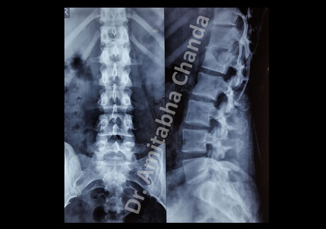
This 20-year-old girl presented with severe pain in back for one month. The pain was more during night. X-ray and MRI showed erosion of L1 vertebral body. Tuberculosis was suspected. CT-guided trucut biopsy confirmed tuberculosis. Minimally Invasive Spinal fixation was done with pedicle screws and rods. She took antitubercular medications for 18 months. She had complete recovery.


Case 02
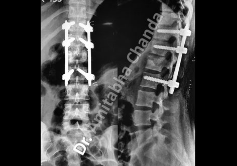
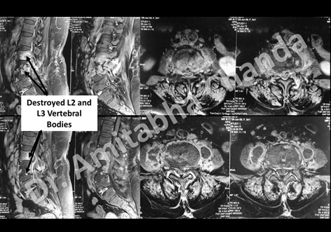
This 54-year-old lady presented with severe back pain for 2 months. The pain increased with time. The pain started radiating to both legs 15 days after the onset of back pain. She was unable to walk or stand for 1 month. She was bedridden for 3 weeks. X-ray lumbar spine showed destruction of the body of L3 vertebra. MRI lumbar spine showed sacralization of L5 vertebra and involvement of L2, L3 and L4 vertebrae with infection (?Tuberculosis). There was pus in the epidural space with compression of neural structures. There was pus in the pre and paravertebral region as well. She was operated. We did a thorough decompression of the neural structures and stabilized the spine with pedicle screws, rods and transverse connectors. She had an excellent recovery. She became ambulatory very soon. The final diagnosis was tuberculosis of spine. Postoperative X-ray looked excellent. She had to take antitubercular medication for 18 months.


